

In addition, ultrasound is sometimes used during surgery by placing a sterile probe into the area being operated on.ĭiagnostic ultrasound can be further sub-divided into anatomical and functional ultrasound. However, to optimize image quality, probes may be placed inside the body via the gastrointestinal tract, vagina, or blood vessels. Most diagnostic ultrasound probes are placed on the skin. Ultrasound probes, called transducers, produce sound waves that have frequencies above the threshold of human hearing (above 20KHz), but most transducers in current use operate at much higher frequencies (in the megahertz (MHz) range). By allowing mothers to see life through ultrasound, we are making it possible for them to choose life!įor more information about A Woman’s Place Medical Clinic, visit For more information about Focus on the Family, visit ultrasound is a non-invasive diagnostic technique used to image inside the body. The ultrasound not only begins that maternal bond but also exhibits that there is a human, with a beating heart, that she helped create. The woman stated, “my boyfriend sleeps like that, I can’t wait to show him.” Being able to see that beautiful image and make a connection while the baby is in the womb is life changing. While scanning a woman just last week, we were able to see the baby’s arms behind her head in the 3D ultrasound. Words no longer hold the same meaning because the abstract images are powerful. Through an ultrasound, it becomes a real baby and the image becomes proof or confirmation of life. When we perform a 3D or 4D ultrasound, it allows the woman to visualize a more defined view of their unborn baby. Ultrasound images are a way to allow women to see life through 2D, 3D, and 4D imaging.

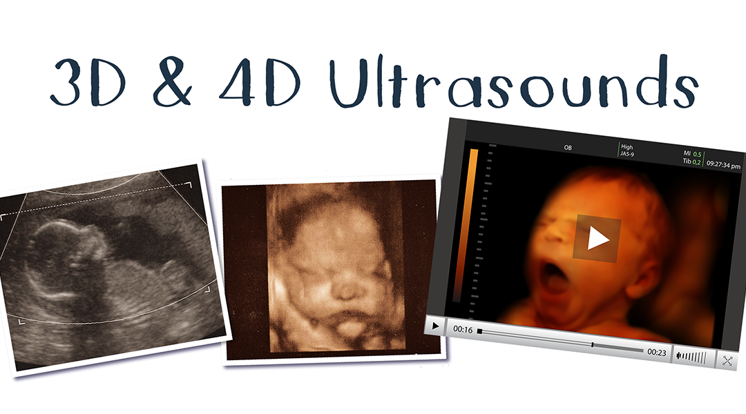
When a woman is faced with the decision of choosing life it is essential for her to see that life with her own eyes. 4D ultrasound brings the static image from a 3D ultrasound to life.Ī woman’s decision whether to keep her baby, place for adoption, or abort is complex and involves many different factors. What is a 4D ultrasound?ĤD ultrasound is different than a 3D ultrasound because it adds the dimension of time, providing a live video of the baby in action: kicking, stretching, yawning, sucking their thumb, opening and closing their eyes. Because of this increased resolution and visibility, 3D ultrasounds capture a more detailed and realistic representation of the developing baby.īecause 3D ultrasounds allow us to view more subtle features, they are helpful in the diagnosis of cleft lip, cleft palate, and heart conditions, and give parents the unique opportunity to see their baby’s face for the very first time.
#3d vs 4d ultrasound series#
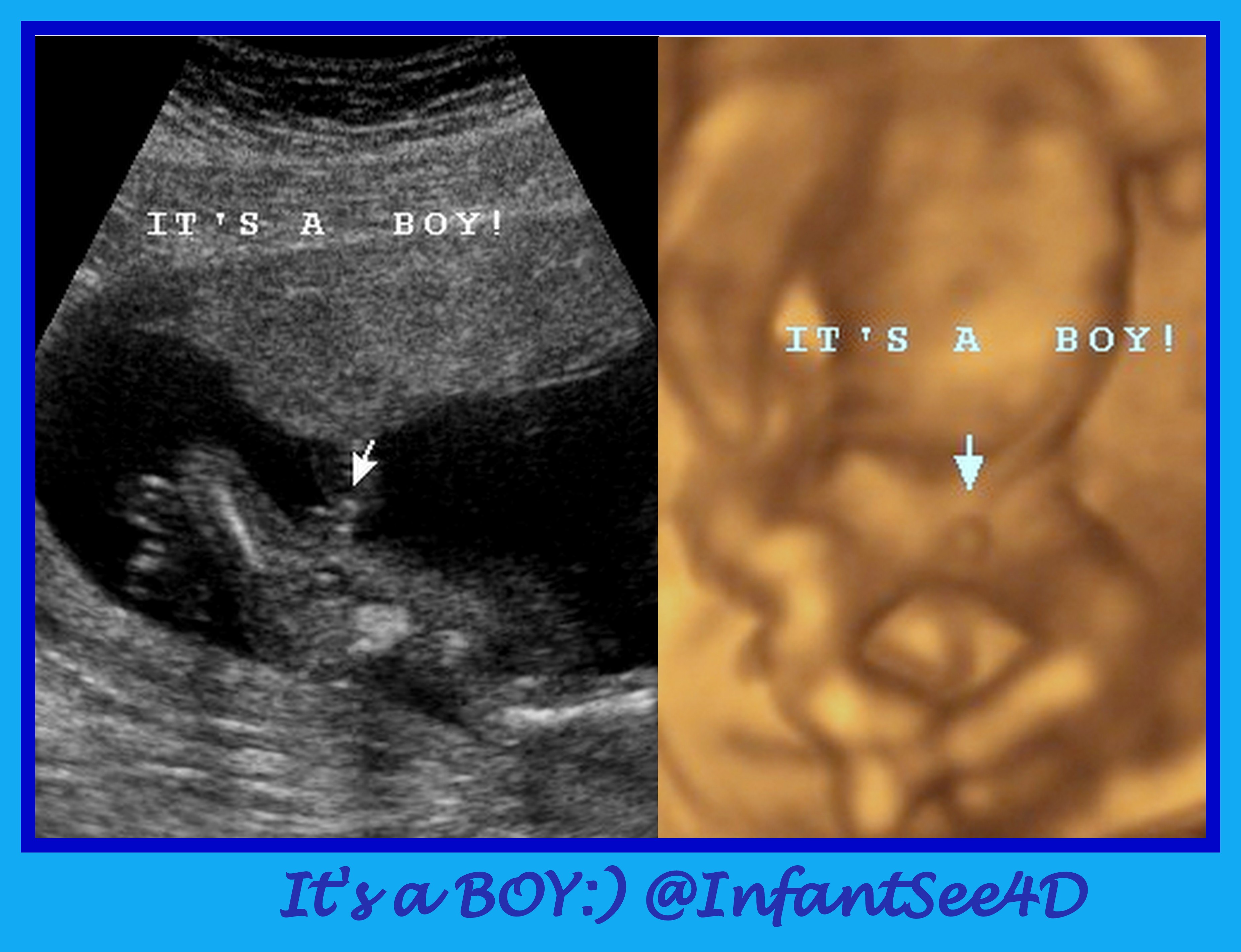
While trained medical professionals have become adept at recognizing features of the developing fetus in 2D, parents are better able to visualize the baby with the advent of 3D ultrasonography.Ī 3D ultrasound creates a different perspective by merging a series of 2D images taken from various angles into a composite to form a 3D picture. This scan is usually repeated at 18-20 weeks of gestation to check for normal growth and development, and to reveal the sex of the baby if desired. Just four weeks after fertilization, the heartbeat can be picked up on an ultrasound. 2D ultrasounds can be done during any trimester, but are often performed early in gestation to confirm the pregnancy and to help establish the due date. The standard 2D obstetric ultrasound shows a flat, black and white picture on a screen. The images can be viewed as pictures on a video screen.” These ultrasound images allow women, men and doctors to have a safe and painless way to see into the womb and see the pre-born baby. The transducer receives these echoes, which are turned into images. According to The American College of Obstetricians and Gynecologists, “The (ultrasound) sound waves come into contact with tissues, body fluids, and bones. Ultrasound scans, also known as sonograms, use the reflection of high-frequency sound waves to create ultrasound images in 2D, 3D and 4D. The following ultrasound information is from that article: What is an ultrasound? In the article “The Differences Between 2D, 3D, and 4D Ultrasounds Explained,” by the Focus on the Family Advocacy team from April 2021, we find an excellent description of the importance of providing ultrasounds to clients in pregnancy medical clinics, like A Woman’s Place Medical Clinics in the Tampa Bay area.


 0 kommentar(er)
